Dura mater
 The cranial meninges consist of three intracranial layers. The dura mater is the outermost layer and is intimately applied to the internal surface of the cranial cavity. The dura mater is comprised of an outer, periosteal, lamina and an inner, meningeal, lamina. The periosteal lamina is applied to the internal surface of the cranial cavity, and the meningeal lamina is a supporting layer that closely reflects the contours of the brain. These two layers of the dura mater separate in certain locations to form dural folds that penetrate the large fissures between parts of the brain. The separation creates spaces that accommodate dural venous sinuses, which receive the venous drainage of the brain. The arachnoid and pia mater are the two layers of the cranial meninges below the dura mater.
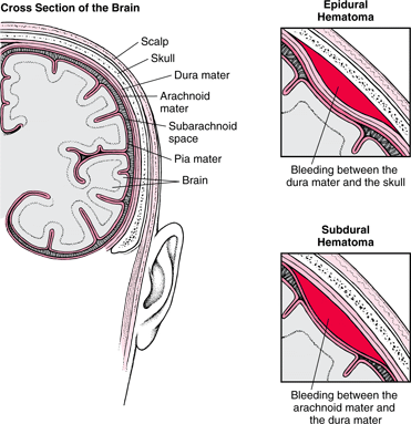
 Extradural or epidural hemorrhage occurs when the meningeal arteries leak blood, and that blood collects between the outer, periosteal, layer of the dura mater, and the skull. This forms an extradural or epidural hematoma. This is usually caused by a hard blow to the head. Typically, a brief concussion occurs, followed by a lucid interval of some hours. Later drowsiness and a coma may occur. As blood mass increases, compression of the brain occurs. The dural border or subdural hematoma results in blood mass that splits open the dural cell border creating a space at the dura-arachnoid mater junction. Dural border hemorrhage is usually venous in origin.
Blood Flow in the Brain
 The brain requires a continuous supply of oxygen and nutrients for function. The cerebral arterial circle, the circle of Willis, is a pentagon shaped circle of vessels on the ventral surface of the brain. The arterial circle is form by the anterior communicating artery, anterior cerebral arteries, internal carotid arteries, posterior communicating arteries, and posterior cerebral arteries. It is an important anastomosis that is located at the base of the brain between the two vertebral and two internal carotid arteries that supply the brain. Blood is supplied to the circle of willis from the basilar artery which divides into the two posterior cerebral arteries, and the internal carotid arteries.
 A stroke is an interruption of the blood supply to any part of the brain. Strokes are the most common neurologic disorder affecting adults in the US. The most common causes of strokes are spontaneous cerebrovascular accidents, such as cerebral thrombosis, cerebral hemorrhage, cerebral embolism, and subarachnoid hemorrhage. An ischemic stroke is generally caused by an embolism in a major cerebral artery. Hemorrhagic stroke follows the rupture of an artery or a sac-like dilation on a weak part of an arterial wall. In time, the weak part of the wall of the aneurysm expands and may rupture, allowing blood to enter the subarachnoid space.
Macula of the Eyeball
The eyeball contains the optical apparatus of the visual system. There are three layers to the eyeball, the fibrous layer, vascular layer, and inner layer. The inner layer of the eyeball is the retina. It is the sensory neural layer of the eyeball. The macula is a small oval area of the retina with special photoreceptor cones that is specialized for acuity of vision. At the center of the macula is the fovea centralis, which is the area of the most acute vision. The macula of the retina is apparent only when the retina is examined with red-free light.
 Age related macular degeneration is a disease that gradually destroys sharp, central vision. As the name suggests, it is associated with aging. The macula is needed for seeing objects clearly and for common daily tasks. There are two forms of age related macular degeneration, wet and dry. In wet age related macular degeneration, abnormal blood vessels behind the retina start to grow under the macula, and these new blood vessels often leak blood and fluid which raises the macula from its normal place. Damage to the macula occurs rapidly. Dry age related macular degeneration occurs when the light-sensitive cells in the macula slowly break down, gradually blurring central vision in the affected eye. Over time, as less of the macula functions, central vision is gradually lost in the affected eye. In some cases, the disease advances slowly and people affected notice little changes in their vision. In other cases, the disease progress faster and may lead to a loss of vision in both eyes. Age related macular degeneration is the leading cause of vision loss in American age 60 and older.
Sources:
Clinically Oriented Anatomy
No comments:
Post a Comment